|
[ Section Lepidella page. ]
[ Amanita Studies home. ]
[ Keys & Checklist/Picturebooks ] "Smith's Lepidella"
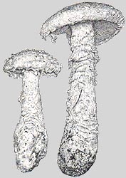
Technical description (t.b.d.) BRIEF DESCRIPTION: The following is based for the most part on the description by Tulloss and Lindgren (1992). The cap of A. smithiana is 50 - 170 mm wide, white to whitish or ivory (with tan, pink, and yellowish tones sometimes present after handling or in dry weather), hemispheric to convex to plano-convex, sometimes somewhat umbonate, with a nonsulcate, appendiculate margin. The flesh is white and unchanging and up to 20 mm thick over the stem. The cap is covered with floccose-felted remnants of volva forming irregularly shaped, floccose warts in young and floccose-felted patches in older specimens. The gills are crowded, free to narrowly adnate, white, bruising slowly to buff or pinkish buff when handled, rarely forking, and 6 - 11 mm broad. The short gills are truncate to subtruncate to rounded truncate to attenuate (the longer ones) and often unevenly distributed among the gills. The stem is about 60 - 180 x 10 - 35 mm, with floccose-felted remnants of the ring near the top and some vague, felted, wart-like remnants of volva at the base of the stem and the top of the bulb. The stipe's bulb is apparently always collected only in part -- (to our knowledge) never with the basal narrow radical (see background silhouette in above illustration on right) in unbroken condition. The large recorded dimensions for a bulb with attached radical are 300 x 55 mm, and the radical in this case was clearly broken at the bottom. The odor can be very unpleasant in age and "truly obnoxious" in senility. At first it is absent to mild to faintly pungent -- described as salty, like dust from Douglas fir bark chips, yeast-like, and like green tomatoes or tomato plants. Persons poisoned by ingestion of A. smithiana described the taste as turnip-like, sometimes with an acidic or bitter component. The spores measure (6.5-) 8.7 - 12.0 (-16.0) x (4.3-) 5.8 - 8.0 (-10.8) µm and are amyloid and ellipsoid to elongate (infrequently broadly ellipsoid to subglobose, infrequently cylindric). Clamps are present at bases of basidia. This species is confirmed as POISONOUS. At least one kidney toxin has been identified in Amanita smithiana. Several cases have been reported in which the kidney or the liver and kidney both ceased to function at least for a time. One of the great dangers is the accidental collection of A. smithiana by commercial harvesters (or private collectors) of one of the American taxa similar to, and sold as, "matsutake" -- Tricholoma magnivelare (Peck) Redhead. It is important for "matsutake" collectors in western North America to be very familiar with A. smithiana and how to distinguish it from the treasured species they are seeking. A 2009 case study and a review of relevant toxicological literature can be downloaded here (PDF 1.33 MB). Poisonings with similar symptomology have been reported for A. nauseosa, A. proxima, A. sphaerobulbosa, and A. thiersii. All species taxonomically related to these taxa should be considered suspect. The species was originally described from Oregon and Washington, U.S.A. The known range now extends from Prov. British Colombia, Canada through the U.S. west of the Great Plains southward to central Mexico. Reports are spotty south of Santa Cruz, California. The species is associated with alder, Douglas Fir (Pseudotsuga), fir, hemlock (Tsuga), larch, pine, and oak. It is probable that A. smithiana occurs throughout the Douglas Fir/Western Hemlock Zone of the Pacific Northwest (Canada and U.S.). This species was often called "A. solitaria" by authors treating fungi of the Pacific Northwest states of the U.S. It is quite distinct from the the European species (A. solitaria (Bull. : Fr.) Fr.). Bas (1969) placed this species in his stirps Rhopalopus. -- R. E. Tulloss and J. E. Lindgren. Drawing (top left): Dr. C. Bas (1969) (NW U.S.A., stipe base radical missing in both fruiting
bodies, reproduced by courtesy of Persoonia, Leiden, the Netherlands)
[ Section Lepidella page. ]
[ Amanita Studies home. ]
[ Keys & Checklist/Picturebooks ] Last changed 22 April 2009. |