
| name | Amanita subnudipes |
| name status | nomen acceptum |
| author | (Romagn.) Tulloss |
| english name | "Undecorated Saffron Ringless Amanita" |
| images |
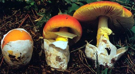
 1. Amanita subnudipes - southwestern France. |
| cap |
The cap of A. subnupides is 30 - 80 mm wide, conic at first, becoming convex, mat, with a striate margin. It is relatively pale orange and slightly more intensely colored over disc. The volva is absent. |
| gills |
The gills are free, subcrowded, and whitish. Short gills are infrequent. |
| stem |
The stem is white or very pale, fragile, exannulate, hollow. The sac-like volva is white, membranous, thin, tall, and persistent. |
| spores | The spores measure (6.2-) 8.9 - 12.2 (-18.1) × (5.5-) 7.5 - 10.4 (-13.2) µm and are subglobose to broadly ellipsoid (rarely globose or ellipsoid or narrower) and inamyloid. Clamps are absent from bases of basidia. |
| discussion | A short key-fragment useful in distinguishing A. crocea (Quél. in Bourd.) Singer exSinger, A. flavescens (E.- J. Gilbert & S. Lund.) Contu, A. romagnesiana Tulloss, and A. subnudipes—as well as the species most phenetically similar to A. fulva (Schaeff.) Fr. is available.—R. E. Tulloss |
| brief editors | RET |
| name | Amanita subnudipes | ||||||||
| author | (Romagn.) Tulloss. 2000. Mycotaxon 75: 329. | ||||||||
| name status | nomen acceptum | ||||||||
| english name | "Undecorated Saffron Ringless Amanita" | ||||||||
| synonyms |
≡Amanita crocea var. subnudipes Romagn. 1982. Bull. Trimestriel Soc. Mycol. France 98(2): 166.
≡Amanitopsis crocea var. subnudipes (Romagn.) Wasser. 1992. Fl. Fung. Ucrainicae, Basidiomycetes, Amanitales: 152. n.v. The editors of this site owe a great debt to Dr. Cornelis Bas whose famous cigar box files of Amanita nomenclatural information gathered over three or more decades were made available to RET for computerization and make up the lion's share of the nomenclatural information presented on this site. | ||||||||
| MycoBank nos. | 467532 | ||||||||
| GenBank nos. |
Due to delays in data processing at GenBank, some accession numbers may lead to unreleased (pending) pages.
These pages will eventually be made live, so try again later.
| ||||||||
| holotypes | in herb. H. Romagnesi (PC) | ||||||||
| revisions | Tulloss, here. | ||||||||
| intro |
The following text may make multiple use of each data field. The field may contain magenta text presenting data from a type study and/or revision of other original material cited in the protolog of the present taxon. Macroscopic descriptions in magenta are a combination of data from the protolog and additional observations made on the exiccata during revision of the cited original material. The same field may also contain black text, which is data from a revision of the present taxon (including non-type material and/or material not cited in the protolog). Paragraphs of black text will be labeled if further subdivision of this text is appropriate. Olive text indicates a specimen that has not been thoroughly examined (for example, for microscopic details) and marks other places in the text where data is missing or uncertain. The following material is derived from the protolog of the present species and from original research of R. E. Tulloss. | ||||||||
| pileus | 30 - 80 mm wide, relatively pale pure orange, slightly more intensely colored over disc, conic at first, becoming convex, mat; context not described; margin short striate; universal veil absent. | ||||||||
| lamellae | free, subcrowded, whitish; lamellulae infrequent or scattered. | ||||||||
| stipe | white or very pale, lacking colored fibrillose squamules characteristic of var. crocea, fragile, sometimes with shaggy upward pointing fibrillose scales; context hollow; exannulate; universal veil as saccate volva, white, membranous, thin, tall, persistent. | ||||||||
| odor/taste | Odorless. Taste not recorded. | ||||||||
| macrochemical tests |
none recorded. | ||||||||
| pileipellis | RET: 75 - 160 µm thick, colorless near surface, otherwise yellow to orangish yellow, fully gelatinized only at surface; filamentous, undifferentiated hyphae 2.5 - 12.8 µm wide, branching, densely packed, dominantly subradially arranged, also with criss-crossing fascicles, occasionally with yellowish subrefractive walls; vascular hyphae 3.8 - 15.4 µm wide, branching, common, sinuous, with irregular outline, locally in complex knots or tangles. | ||||||||
| pileus context | RET: filamentous, undifferentiated hyphae 1.8 - 11.0 µm wide, branching, plentiful, in fascicles, with fascicles forming loosely interwoven lattice; acrophysalides badly collapsed and broken, clavate, up to 78 × 35 µm and probably larger, plentiful(?), wall thickness not assessable; vascular hyphae 4.0 - 19.0 µm wide, branching, relatively common, with frequent abrupt constrictions. | ||||||||
| general context | double click in markup mode to edit. | ||||||||
| lamella trama | RET: bilateral; wcs = 50 - 55 µm; subhymenial base shallow, dominated by filamentous, undifferentiated hyphae and narrow inflated elements (subglobose to broadly clavate to clavate to subcylindric to fusiform, sometimes in chains, up to 47 × 14.1 µm and as small as 5* µm major diameter, sometimes penetrating subhymenium) diverging at shallow angle or at an angle up to 45° to central stratum; central stratum including intercalary slightly inflated narrowly fusiform cells (e.g., 55 × 15.0 µm); filamentous, undifferentiated hyphae µm, 2.8 - 8.5 µm, branching; terminal, divergent inflated cells not observed; vascular hyphae 4.0 - 6.3 µm wide, infrequent, serpentine, occasionally with tight coils, apparently restricted to the central stratum. | ||||||||
| subhymenium | RET: wst-near = (35-) 50 - 65 µm; wst-far = 60 - 85 µm; branching structure of uninflated and partially inflated and branched hyphal segments and small inflated cells (sometimes dominating), with occasional hyphal segments running subparallel to central stratum, with basidia arising from cells of all types and rarely from elongate element of subhymenial base. | ||||||||
| basidia | 41 - 71 × 10.0 - 15.8 µm, dominantly 4-, rarely 1-sterigmate, with sterigmata up to 6.5 × 4.8 µm; clamps not observed. | ||||||||
| universal veil | RET: On pileus of mature basidiome: over much of surface as scattered gelatinized fragments consistent with structure of interior of limb on stipe base [e.g., collapsed (flattened) and gelatinized cell 140 × 102 µm]. On pileus of incompletely basidiome: scattered small patches or tufts of tangled, frequently branching, filamentous, undifferentiated hyphae, collapsed, partially gelatinized here and there, with clavate terminal cells up to 40 × 16.3 µm. On stipe base, exterior surface: substantial layer several hyphal diameters thick; filamentous, undifferentiated hyphae 3.2 - 10.0 µm wide, branching, many with sublongitudinal orientation, singly and in narrow fascicles forming loosely interwoven lattice, almost all collapsed; vascular hyphae 5.5 - 19.5 µm wide, infrequent, infrequently branching, fragmented, partially gelatinized in spots. On stipe base, interior: filamentous, undifferentiated hyphae 2.0 - 12.8 µm wide, branching, dominant, singly and in often rather broad fascicles forming open lattice; inflated cells scattered, very infrequent, subglobose to broadly clavate to clavate, up to 120* × 52 µm; vascular hyphae 4.2 - 14.0 µm wide, infrequent. On stipe base, inner surface: in some regions like interior but partially gelatinized, other regions comprising very thin layer of sublongitudinally oriented filamentous, undifferentiated hyphae; inflated cells on surface collapsed, scattered, infrequent, up to 141 × 50 µm; inflated cells free in mount gelatinized, numerous, yellowish or orangish, possibly(?) from friable part of limbus internus or remains of layer of inflated cells from lamella edge on limbus internus surface. | ||||||||
| stipe context | RET: longitudinally acrophysalidic; filamentous, undifferentiated hyphae 3.5 - 10.2 µm wide, branching, plentiful; acrophysalides dominant, thin-walled, up to 298 × 39 µm; vascular hyphae 3.8 - 9.8 µm wide, scattered, infrequently branching, sinuous, locally with numerous abrupt constrictions, locally in coils and tangles. | ||||||||
| lamella edge tissue | RET: frequent and extended remains of layer up to 56 µm thick; cells densely packed, in 3 - 6 layers, up to 41 × 36 µm, partially gelatinized and collapsed, with few to plentiful strands of filamentous, undifferentiated hyphae 4.6 - 5.2 µm wide, radially oriented, partially gelatinized. | ||||||||
| basidiospores | RET: [145/5/4] (6.2-) 8.9 - 12.2 (-18.1) × (5.5-) 7.5 - 10.5 (-13.2) µm, (L = 9.5 - 11.2 µm; L’ = 10.6 µm; W = 8.5 - 9.8 µm; W’ = 9.2; Q = (1.03-) 1.05 - 1.29 (-2.29); Q = 1.12 - 1.16 (-1.22); Q’ = 1.15), hyaline, colorless, smooth, thin-walled, inamyloid, subglobose to broadly ellipsoid, occasionally ellipsoid, rarely globose, rarely elongate to cylindric, occasionally langeniform to clavate in exsiccata early in sporulation when collected and dried, adaxially flattened, occasionally expanded at one end; apiculus sublateral, cylindric; contents granular to multi- or monoguttulate; white in deposit. | ||||||||
| material examined | RET: FRANCE: CORSICA—Galeria, valley of R. Fango, 3.xi.1982 R. A. Maas Geesteranus 15720 (L). OISE—Apremont, 27.vii.1953 H. Romagnesi 53.114 (holotype, in herb. H. Romagnesi => PC). ITALY: SARDINIA—Cantoniera Catala, ca. Calangianus, 1.xi.1983 M. Moser 83/515 (IB). TRENTO—Pergine Valsugana, Rifugio Alpino Marzola, 31.vii.1995 Marco Floriani 177 (in herb. Floriani; RET 257-7). UNKN.—unkn. loc., 3.x.2010 Carmine Lavorato 101003-10 (RET 502-1, nrITS seq'd.). | ||||||||
| citations | —R. E. Tulloss | ||||||||
| editors | RET | ||||||||
Information to support the viewer in reading the content of "technical" tabs can be found here.
Each spore data set is intended to comprise a set of measurements from a single specimen made by a single observer; and explanations prepared for this site talk about specimen-observer pairs associated with each data set. Combining more data into a single data set is non-optimal because it obscures observer differences (which may be valuable for instructional purposes, for example) and may obscure instances in which a single collection inadvertently contains a mixture of taxa.


Text and User-Generated Sporographs are published under the Creative Commons License.

In the case of a taxon page, image credits are on the 'image' tab.