| name |
Amanita sp-S07 |
| author |
Tulloss |
| name status |
cryptonomen temporarium |
| GenBank nos. |
Due to delays in data processing at GenBank, some accession numbers may lead to unreleased (pending) pages.
These pages will eventually be made live, so try again later.
|
| intro |
Olive text indicates a specimen that has not been
thoroughly examined (for example, for microscopic details) and marks other places in the text
where data is missing or uncertain.
The following material is based on original research by R. E. Tulloss. |
| pileus |
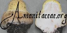
88 mm wide, yellowish-brown with greenish or grayish tint, campanulate to planoconvex, dry, dull; context white, unchanging when cut or bruised, 7 mm thick above stipe, thinning evenly to margin; margin striate (0.15 - 0.2R), nonappendiculate; universal veil absent. |
| lamellae |
free, without decurrent line on stipe, crowded, sordid tan in mass, sordid pale tan in side view, unchanging when cut or bruised, 5 mm broad; lamellulae truncate. |
| stipe |
215 × 14 mm, whitish, narrowing upward, surface fibrillose and longitudinally striate; context whitish, apparently unchanging when cut or bruised, stuffed with white cottony material, with 6 mm wide central cylinder; exannulate; universal veil as saccate volva, white, leathery and rather tough, 39 mm from base to highest point of limb. |
| odor/taste |
Odor and taste not recorded (single specimen somewhat senescent). |
macrochemical
tests |
none recorded. |
| lamella trama |
bilateral; wcs = 55 - 60 µm; dominated by subhymenial base; filamentous, undifferentiated hyphae ?? µm wide, branching, ??, commonly giving rise to elements of subhymenium; divergent inflated cells plentiful (ovoid to broadly clavate to clavate to subcylindric, up to 65 × 44 µm, with walls up to 1.0 µm thick, some in short chains, many intercalary between uninflated hyphal segments, many terminal, many of latter rostrate); vascular hyphae ?? µm wide, ??. |
| subhymenium |
wst-near = 80± µm; wst-far = 110 - 115 µm; dominantly cellular (1 - 2 cells deep), with basidia arising from uninflated and partially inflated hyphal segments as well as inflated cells. |
| basidia |
35 - 64 × 11.0 - 14.5 µm, 4- and (occasionally) 1-sterigmate; clamps not observed. |
| partial veil |
absent. |
| lamella edge tissue |
sterile. |
| basidiospores |
[40/1/1] (9.0-) 9.2 - 12.5 (-15.5) × (6.5-) 6.8 - 9.2 (-11.8) µm, L = 10.1 µm; W = 7.9 µm; Q = (1.06-) 1.08 - 1.60 (-1.86); Q = 1.26), hyaline, colorless, smooth, thin-walled, inamyloid, subglobose to broadly ellipsoid to ellipsoid to elongate, usually at least somewhat adaxially flattened; apiculus sublateral, cylindric; contents monoguttulate; ?? in deposit. |
| ecology |
Solitary in loam over red clay In mixed deciduous/coniferous woods near Pinus sp. and Quercus muehlenbergii, under dry conditions, but with many large boletes and amanitas fruiting and in good condition. |
| material examined |
U.S.A.: SOUTH CAROLINA—Greenville Co. - Roper Mountain Park, Emily & Erin Luetkemeier & R. E. Tulloss 8-23-91-A (RET 032-3). |
| discussion |
Some spores of Tulloss 8-23-91-A were slightly malformed and rather large.
The following sentence transcribed from my notes dated 12.ix.1991 suggests to me (at present) that the "rostrate" appearance of "terminal" inflated cells of the lamella trama may indicate that they were not terminal, but that breakage of hyphae during sectioning may have made them appear to be both terminal and rosrate: "On those terminal inflated cells of the lamella trama that are rostrate, the narrow apical process is often curved or, in some other manner, suggestive that it would have progressed to give rise to an uninflated hyphal segment." |
| citations |
—R. E. Tulloss |
| editors |
RET |
|


