
| name | Amanita pallidocarnea | ||||||||
| author | ("pallido-carnea") (Höhn.) Boedijn. 1951. Sydowia 5: 319. | ||||||||
| name status | nomen acceptum | ||||||||
| english name | "Pale Flesh-Colored Ringless Amanita" | ||||||||
| synonyms |
≡Amanitopsis vaginata var. pallidocarnea ("pallido-carnea") Höhn. 1914. Sitzb. Akad. Wiss. Wien, Math. Nat. Kl. Abt. I. 123: 74. The editors of this site owe a great debt to Dr. Cornelis Bas whose famous cigar box files of Amanita nomenclatural information gathered over three or more decades were made available to RET for computerization and make up the lion's share of the nomenclatural information presented on this site. | ||||||||
| MycoBank nos. | 292457, 479209 | ||||||||
| GenBank nos. |
Due to delays in data processing at GenBank, some accession numbers may lead to unreleased (pending) pages.
These pages will eventually be made live, so try again later.
| ||||||||
| lectotypes | in herb. F. von Höhnel (FH) | ||||||||
| lectotypifications | Z. L. Yang. 2001. Mycotaxon 80: 281, figs. 1-5. | ||||||||
| type studies | Z. L. Yang. 2001. Mycotaxon 80: 281, figs. 1-5. | ||||||||
| revisions | Z. L. Yang. 2001. Mycotaxon 80: 281, figs. 1-5. | ||||||||
| intro |
The following text may make multiple use of each data field. The field may contain magenta text presenting data from a type study and/or revision of other original material cited in the protolog of the present taxon. Macroscopic descriptions in magenta are a combination of data from the protolog and additional observations made on the exiccata during revision of the cited original material. The same field may also contain black text, which is data from a revision of the present taxon (including non-type material and/or material not cited in the protolog). Paragraphs of black text will be labeled if further subdivision of this text is appropriate. Olive text indicates a specimen that has not been thoroughly examined (for example, for microscopic details) and marks other places in the text where data is missing or uncertain. The following material is based entirely on (Yang 2001). Basidiome small to medium-sized. | ||||||||
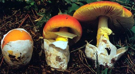
| pileus | 40 - 60 mm wide, convex to applanate, slightly umbonate, dark grey to dark brown over disc [fuscous to fuscous-black to Benzo brown; 5E4, 6E4], becoming gray to grayish brown [hair brown to drab to cinnamon-drab; 6D3, 6D4] toward the margin; context white; margin striate (0.3 - 0.5R), non-appendiculate; universal veil absent or sometimes as small, felted to submembranous, dirty white, fragments. | ||||||||
| lamellae | free, crowded, pink [flesh pink; 8A2, 8A3, 9A2, 9A3], up to 5 mm broad; lamellulae truncate to subtruncate, in 1 - 2(-3) ranks, evenly distributed. | ||||||||
| stipe | 60 - 100 × 5 - 15 mm, subcylindric or slightly tapering upwards, with apex slightly expanded, pinkish, below apical region covered with fine pink [flesh pink to orient pink; 9A2, 8A2] squamules, becoming fibrillose and paler downwards, with apical region farinose and pinkish to pink; context whitish, hollow; exannulate; universal veil saccate, thin, membranous, 20 - 40 × 15 - 25 mm, with both surfaces dirty white. | ||||||||
| odor/taste | not recorded. | ||||||||
| macrochemical tests |
none recorded. | ||||||||
| pileipellis | 50 - 80 µm thick; suprapellis 20 - 30 µm thick, strongly gelatinized, comprising subradially arranged, filamentous hyphae 2 - 4 µm wide, thin-walled, subhyaline or sometimes with brownish vacuolar pigments; subpellis 30 - 50 µm thick, composed of subradially and compactly arranged, filamentous hyphae 5 - 12 µm wide, with brownish to brown, vacuolar pigments; vascular hyphae rare, 3 - 10 µm wide. | ||||||||
| pileus context | not described. | ||||||||
| lamella trama | poorly rehydrating in specimens studied. | ||||||||
| subhymenium | 30 - 40 (-50) µm thick, with 2 - 3 layers of subglobose to ovoid or doliform, inflated cells, 12 - 20 × 10 - 15 µm, mixed with hardly inflated hyphal segments, 4 - 7 µm wide. | ||||||||
| basidia | 40 - 70 (-80) × 12 - 18 µm, 4-sterigmate, with sterigmata 4 - 6 µm long; clamps absent. | ||||||||
| universal veil | On pileus, excluding outer surface: composed of (very) abundant filamentous hyphae 2 - 8 µm wide, colorless hyaline, thin-walled, frequently branching, interwoven; inflated cells fairly abundant to abundant, subglobose to ovoid (60 - 100 × 50 - 80 µm) sometimes ellipsoid (65 - 100 × 20 - 50 µm) or sphaeropedunculate (60 - 70 × 40 - 5 µm), usually single and terminal, colorless hyaline, thin-walled; vascular hyphae rare, 3 - 12 µm wide. On pileus, outer surface: usually with more abundant filamentous hyphae than in interior. On stipe base, interior: composed of abundant to very abundant filamentous hyphae, 3 - 7 µm wide, colorless hyaline, thin-walled, branching and interwoven, sometimes anastomosing; inflated cells fairly abundant to abundant, subglobose to ovoid (30 - 70 × 25 - 60 µm) or ellipsoid (50 - 80 × 20 - 50 µm), hyaline, colorless or occasionally with brownish to yellowish contents, thin-walled, terminal, usually single, sometimes in chains of 2 - 3; vascular hyphae rare to scattered to locally conspicuous, 2 - 6 µm wide. On stipe base, outer surface: similar to the interior, but with more abundant filamentous hyphae. On stipe base, inner surface: strongly gelatinized, composed of filamentous hyphae 2 - 5 µm wide. | ||||||||
| stipe context | longitudinally acrophysalidic; acrophysalides 180 - 280 × 25 - 45 µm; filamentous hyphae 2 - 8 µm wide, scattered in interior, fairly abundant on stipe surface; vascular hyphae rare, 3 - 20 µm wide. | ||||||||
| lamella edge tissue | as sterile, somewhat gelatinized strip up to 200 µm wide in side view, composed of very abundant filamentous hyphae, 3 - 7 µm wide, hyaline, colorless or with yellowish contents, subparallel to lamella edge, mixed with abundant inflated cells, ovoid to subglobose (30 - 50 × 25 - 40 µm) to broadly ellipsoid (35 - 60 x 20 - 35 µm) or broadly clavate (40 - 55 × 20 - 25 µm), thin-walled, hyaline, colorless or sometimes with yellowish contents, terminal, single or in chains of 2 - 3; vascular hyphae rare. | ||||||||
| basidiospores |
from type study by Yang (2001: 284): [30/1/1] ((9.0-) 9.5 - 12.5 (-14.5) × 8.5 - 11.5 (-14.0) μm, ( Composite of all material revised by Yang (2001): [75/3/2] (8.5-) 9.0 - 12.0 (-14.5) × 8.0 - 11.0 (-14.0) μm, ( | ||||||||
| ecology | from Yang (2001): On the ground, in broad-leaved forests. | ||||||||
| material examined |
from Yang (2001):
CHINA:
HAINAN—Ledong Li Autonomous Co. - Jianfengling, 20.viii.1999 M. S. Yuan 4373 (HKAS 34571). Changjiang Li Autonomous Co. - Bawangling, 27.vi.1995 X. L. Wu s.n. (HKAS 36701, two colored photos, no preserved exsiccata).
INDONESIA: JAVA—Tjibodas, | ||||||||
| discussion |
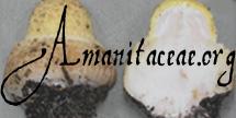
Unfortunately, the lectotype of A. pallidocarnea comprises only an imperfect fruitbody—about a quarter of the pileus connecting with the upper part of the stipe; the lower part of the stipe, and the volva on the stipe base are not preserved. A felty to submembranous, dirty white, volval remnant was found on the pileus (fig. 1). Due to its basidiome with pink lamellae, and a saccate volva on the stipe base, features shared with the genus Volvariella, A. pallidocarnea was suspected of not being a member of the genus Amanita (Yang 1997: 102). Having studied the lectotype, and additional material, I found that it is an Amanita, and is characterized by its dark colored pileus with relative long striations along the pileal margin, its pink lamellae, pinkish stipe, globose to subglobose spores, and volval remnants with fairly abundant to abundant inflated cells. Amanita vaginata var. roseilamellata Bresinsky, which was originally described from Germany, also has pinkish lamellae (Bresinsky & Einhellinger, 1987), but differs from A. pallidocarnea by its paler pileus, white to greyish stipe, and narrower spores. Furthermore, judging from the description and illustrations provided by Bresinsky & Einhellinger (1987), A. vaginata var. roseilamellata might have shorter striations along the pileal margin and a more strongly constructed volva with fewer inflated cells. | ||||||||
| citations | —Zhu L. Yang | ||||||||
| editors | RET | ||||||||
Information to support the viewer in reading the content of "technical" tabs can be found here.
Each spore data set is intended to comprise a set of measurements from a single specimen made by a single observer; and explanations prepared for this site talk about specimen-observer pairs associated with each data set. Combining more data into a single data set is non-optimal because it obscures observer differences (which may be valuable for instructional purposes, for example) and may obscure instances in which a single collection inadvertently contains a mixture of taxa.


Text and User-Generated Sporographs are published under the Creative Commons License.

In the case of a taxon page, image credits are on the 'image' tab.
