
| name | Amanita mira |
| name status | nomen acceptum |
| author | Corner & Bas |
| english name | "Gold Coin Amanita" |
| images | |
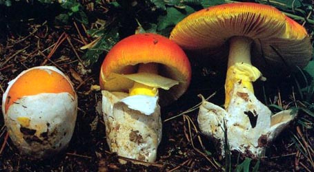
| cap | Much of this information is taken from Corner and Bas (1962). The cap of A. mira is 40 - 90 mm wide, campanulate to plane with a depressed center, subviscid, with a finely tuberculate-striate margin. The cap is orange-red to pale clear orange in the center, yellow orange, ochre-yellow, or bright yellow toward pale margin. The cap is sprinkled with small, firm, yellowish to whitish, pyramidal warts. |
| gills | The gills are free, crowded, thin, and white. |
| stem | The stem is 50 - 110 × 5 - 9 mm, equal or tapering upward, solid, becoming hollow, white or slightly grayish, mostly exannulate, with 2 - 3 more or less complete rings of small, subfloccose, yellow warts at the base (as on the cap). |
| spores | According to Corner & Bas (1962), the spores measure 6.4 - 7.9 × 6.2 - 7.7 µm when dried (7.0 - 8.5 × 6.5 - 7.5 µm, fresh) and are globose to subglobose (rarely broadly ellipsoid) and inamyloid. Clamps are not found at bases of basidia. |
| discussion |
Corner records that monkeys ate this species "without discomfort." Described from forest in Singapore. It has also reported from China (Yunnan Prov. according to Yang(1997)), and Malaya.—R. E. Tulloss |
| brief editors | RET |
| name | Amanita mira | ||||||||
| author | Corner & Bas. 1962. Persooni 2: 290, pl. 9c, figs. 48-50. | ||||||||
| name status | nomen acceptum | ||||||||
| english name | "Gold Coin Amanita" | ||||||||
| MycoBank nos. | 282979 | ||||||||
| GenBank nos. |
Due to delays in data processing at GenBank, some accession numbers may lead to unreleased (pending) pages.
These pages will eventually be made live, so try again later.
| ||||||||
| holotypes | L | ||||||||
| revisions | Z. L. Yang. 1997. Biblioth. Mycol. 170: 32, figs. 19-20. | ||||||||
| intro |
The following text may make multiple use of each data field. The field may contain magenta text presenting data from a type study and/or revision of other original material cited in the protolog of the present taxon. Macroscopic descriptions in magenta are a combination of data from the protolog and additional observations made on the exiccata during revision of the cited original material. The same field may also contain black text, which is data from a revision of the present taxon (including non-type material and/or material not cited in the protolog). Paragraphs of black text will be labeled if further subdivision of this text is appropriate. Olive text indicates a specimen that has not been thoroughly examined (for example, for microscopic details) and marks other places in the text where data is missing or uncertain. NOTE: Spore data from papers by Z. L. Yang are presented following his use of the "Times New Roman" face for "Q" and "Q'"—respectively, " | ||||||||
| pileus | from protolog: 40 - 90 mm wide, orange-red to pale clear orange in center, yellow-orange, ochre-yellow, or bright yellow toward pale margin, generally becoming dingy fuliginous olive or bistre from center outward to margin with age, campanulate to plane with depressed center, subviscid; context white except for yellowish below pileipellis, 3 - 5 mm thick above stipe, membranous in limb; margin finely tuberculate-striate (0.5R); universal veil as sprinkling of small firm yellowish to whitish pyramidal warts about 1 mm high and 1 - 2 mm wide, often glabrous after rain. | ||||||||
| lamellae | from protolog: free, crowded, white, thin, 4 - 10 mm broad, with 80 - 100 primaries, with edges entire; lamellulae truncate, with 0 - 1 between each pair of otherwise adjacent lamellae. | ||||||||
| stipe | from protolog: 50 - 100 × 5 - 9 mm, white or slightly graying, finely appressed fibrillose, cylindric or narrowing upward; bulb 8 - 15 mm wide, proportionately small; context solid, becoming hollow; partial veil seen only once as distinct pendant collapsed ring at stipe apex, mostly exannulate; universal veil as 2 - 3 more or less complete rings of small sub-floccose yellow warts (as on pileus) on upper part of bulb or as yellow floccose-felted slightly warty coating of bulb. | ||||||||
| odor/taste | not recorded. | ||||||||
| macrochemical tests |
none recorded. | ||||||||
| pileipellis | from protolog: about 150 µm thick, without hyaline gelatinous suprapellis; filamentous hyphae 3 - 7 µm wide, repent, agglutinated, intermixed over disc, wavy-radially oriented near margin, with vacuolar yellow to umber pigment, with scattered repent, rounded terminal segments of hyphae. | ||||||||
| pileus context | not described. | ||||||||
| lamella trama | from protolog: hardly analyzable in dried material; with many large inflated cells (e.g., 65 × 35 μm, 80 × 50 μm, 125 × 50 μm). | ||||||||
| subhymenium | not described. | ||||||||
| basidia | from protolog: 30 - 40 × 10 - 13 um, 4-sterigmate, with sterigmata about 4 µm long. | ||||||||
| universal veil | from protolog: On pileus: with elements having more or less anticlinal orientation; filamentous hyphae 4 - 8 μm wide; inflated cells in erect chains, narrowly to broadly cylindric or ellipsoid or clavate, 27 - 72 × 7 - 40 um, with apical cells often more or less acuminate. On stipe base: filamentous hyphae present; inflated cells ellipsoid or ovoid or clavate, up to 40 × 30 μm. | ||||||||
| stipe context | from protolog: hardly analyzable in dried material; longitudinally acrophysalidic; acrophysalides up to 40 µm wide and apparently up to 150 µm long and longer, with many twisting refractive hardly septate hyphae up to 25 µm wide. No clamps observed. | ||||||||
| partial veil | absent. | ||||||||
| lamella edge tissue | from protolog: inflated cells 15 - 35 × 5 - 15 um, cylindric, clavate or pyriform, colorless, thin-walled, smooth, forming sterile lamellae edge. | ||||||||
| basidiospores |
from protolog: [-/-/-] 6.4 - 7.9 × 6.2 - 7.7 μm, (Q = 1.0 - 1.1), colorless, hyaline, thin-walled, globose to subglobose; apiculus not described; contents "with one large gutta or several small ones"; color in deposit not reported. from revision by Yang (1997) [190/8/6] 6.0 - 8.0 (-9.0) × (5.0-) 6.0 - 7.5 (-8.0) μm, ( | ||||||||
| ecology | Solitary or in small groups. China: At 580-1000 m elev. In forests under Castanopsis and Lithocarpus. Singapore: Common in forest every rainy season. | ||||||||
| material examined |
from protolog: SINGAPORE: Botanical Garden, 15.ix.1940 E. J. H. Corner s.n. (L, watercolor drawing, as "Amanitopsis 4"); Bukit Timah, 16.viii.1939 E. J. H. Corner s.n. (holotype, L), 21.viii.1939 E. J. H. Corner s.n. (paratype, ?L). from Yang (1997): CHINA: YUNNAN—Xishuangbanna Dai Autonomous Prefecture - Mengla Co., Menglun, Botanical Garden, 12.viii.1988 Zhu L. Yang 376 (HKAS 21776); Mengla Co., Menglun, Mangangshan, 31.x.1989 Zhu L. Yang 886 (HKAS 22549); Mengla Co., Menglun, 600 m elev., 11.ix.1974 S. Y. Zeng. s.m. (HKAS 1458a), 30.x.1989 Zhu L. Yang 867 (HKAS 22551); Mengla Co., Menglun, 650 m elev., 9.viii. Zhu L. Yang 1493 (HKAS 24170); Mengla Co., Menglun, 1000 m elev., 12.viii.1995 Zhu L. Yang 2163 (HKAS 29526). | ||||||||
| discussion |
from protolog: "This species bears a certain resemblance to Amanita muscaria..., but is easily distinguished by the smaller and globulose spores, by the different structure of the remnants of the volva on the pileus which results in small firm pyramidal warts, and by the generally lacking annulus. Amanita muscaria has never been observed in Malaya." The lack of a gelatinized suprapellis and the form of the warts on the pileus and stipe bulb suggest this species is more closely related to A. farinosa than to the clamp-bearing muscarioid taxa. While presence/absence of clamps was not reported in the protolog (probably due to the condition of the tissue in the exsiccata), clamps are probably absent given the similarity to A. farinosa. Colors reported for the center of the pileus differ in the two regions for which reports have been made. | ||||||||
| citations | —R. E. Tulloss | ||||||||
| editors | RET | ||||||||
Information to support the viewer in reading the content of "technical" tabs can be found here.
| name | Amanita mira |
| name status | nomen acceptum |
| author | Corner & Bas |
| english name | "Gold Coin Amanita" |
| images | |
| watercolor | Prof. E. J. H. Corner (1-4) Singapore, illustrations from original description (Corner and Bas, 1962) reproduced by courtesy of Persoonia, Leiden, the Netherlands. |
Each spore data set is intended to comprise a set of measurements from a single specimen made by a single observer; and explanations prepared for this site talk about specimen-observer pairs associated with each data set. Combining more data into a single data set is non-optimal because it obscures observer differences (which may be valuable for instructional purposes, for example) and may obscure instances in which a single collection inadvertently contains a mixture of taxa.


