
| name | Amanita infusca sensu Pegler and Shah-Sm. | ||||||||||||
| name status | sensu | ||||||||||||
| GenBank nos. |
Due to delays in data processing at GenBank, some accession numbers may lead to unreleased (pending) pages.
These pages will eventually be made live, so try again later.
| ||||||||||||
| intro |
Olive text indicates a specimen
that has not been
thoroughly examined (for example, for microscopic
details) and marks other places in the text
where data is missing or uncertain. Macroscopic characters of fresh material are derived largely from (Pegler and Shah-Smith 1997). The macroscopic description of (Tang et al. 2015) conflicts on a number of points, and these conflicts are addressed in editorial notes. Microscopic characters are derived both from the Pegler and Shah-Smith description and from Tang et al. Again, apparent conflicts are addressed in editorial notes. The Pegler and Shah-Smith description was published approximately two years after the material was collected. The Tang et al description was published 20 years after the original collection. When Tang et al. differ by using part of the description from the protolog (e.g., for color information) we give greater credence in such cases to Pegler and Shah-Smith, the latter of whom was the collector of the material. NOTE: Spore measurements from papers by Z. L. Yang use his "Times New Roman" face for "Q" and "Q'"—respectively, " Tang et al. (2015): Basidiome medium to large. | ||||||||||||
| pileus |
Pegler and
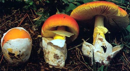
Shah-Smith (1997): 70 - 140 mm wide, dark reddish brown, darker at
margin, sometimes paling
to yellowish brown over disc, broadly convex to
planar, finally with margin upturned, viscid;
context not described; margin
short [per figure] plicate-striate, nonappendiculate;
universal veil absent [per figure]. [Note: Tang et al. (2015) differ in citing (exclusively) the pileus colors and shape from the protolog of Amanita infusca. They plausibly estimate the marginal striations to occupy 10% of the pileus radius.—ed.] | ||||||||||||
| lamellae |
Pegler and
Shah-Smith (1997): free, crowded, white, up to 10 mm
broad, with edge
white or brown; lamellulae truncate, with at
least two lengths. [Note: Tang et al. (2015) report the lamellae as pale brown with "umbrine edge." This appears to be a report on colors in the dried material.—ed.] | ||||||||||||
| stipe |
Pegler and
Shah-Smith (1997): 80 - 125 × 10 - 20 mm, pale brown
cylindric or narrowing upward, decorated with pale
brown squamules above partial veil and with reddish
brown to dark reddish brown squamules below partial
veil; context stuffed;
partial veil subapical, membranous,
persistent, pale to reddish brown;
universal veil as saccate volva, firmly
fleshy, white with brownish gray edge, remaining
entirely within substrate. [Note: The difference between the two cited paper with regard to the stipe can be attributed to the Tang et al. (2015) description's having been based on dried material. The most important differences (because of their impact on the two reports on microscopic anatomy) are the differences in description of the saccate universal veil. Unlike the "white with brownish gray margin" of Pegler and Shah's description, Tang et al. describe the volva as "the upper half grey-black, the lower half greyish white." Pegler and Shah-Smith report finding hyaline tissue including scattered inflated cells in the volval tissue. They do not describe the hyphae has being pigmented or as having notably frequent septa or as sometimes having walls that would be very thick for an Amanita. The Pegler and Shah-Smith description is compatible with the tissue of the interior of most fleshy, saccate volvas in sections Caesareae and Vaginatae. Apparently some deterioration of the specimen (possibly due in part to attack by a brown, thick-walled hyphomycete) occurred between the review of the material by Pegler and the review of the material by Tang. Occasionally one sees evidence of such foreign tissue on or near surfaces of exsiccata of amanitas.—ed.] | ||||||||||||
| odor/taste | none. | ||||||||||||
| macrochemical tests |
none recorded. | ||||||||||||
| pileipellis |
Pegler and Shah-Sm. (1997): [including] gelatinized layer of
"firmly agglutinated" hyphae; filamentous hyphae
1.5 - 4.0 μm wide, interwoven, with thin brown
wall. [Note: While the tissue is described as an ixocutis, the mention of brown cell walls implies an ungelatinized layer of hyphae is also present. A pileipellis with two distinct layers is the dominant form in the genus Amanita. So far as is known to the editor, this is the only form that pileipellis takes in sect. Caesareae.—ed.] | ||||||||||||
| pileus context | not described in either reference. | ||||||||||||
| lamella trama | Pegler and Shah-Sm. (1997): bilateral, divergent; filamentous hyphae 3.0 - 5.0 μm wide, with intercalary inflated segiments up to 20 μm wide; clamps present. | ||||||||||||
| subhymenium | Pegler and Shah-Sm. (1997): pseudoparenchymatous; 17.0 - 26 μm wide. | ||||||||||||
| basidia |
Pegler and
Shah-Smith (1997): 45 - 55 × 10.0 - 13.0 μm,
4-sterigmate; clamps present at base. Tang et al. (2015): 55 - 65 × 7.0 - 11.0 μm, 4-sterigmate, with sterigmata up to 4.0 - 5.0 μm long; clamps abundant. | ||||||||||||
| universal veil |
Pegler and Shah-Sm. (1997): On stipe base: filamentous
hyphae dominating, 2.0 - 9.0 μm wide, loosely
interwoven, hyaline; inflated cells scattered,
globose to clavate, 15. - 45 μm wide; clamps
abundant. Tang et al. (2015): On stipe base: filamentous hyphae dominant and densely arranged, 5.0 - 10.0 μm wide, frequently septate, often with brownish vacuolar pigment, with walls thin or up to 1.0 - 2.0 μm thick; inflated cells rare; vascular hyphae rare; clamps abundant. [Note: There are marked differences in these two descriptions. And these can be related to differences in description of the unversal veil in the two cited articles. See the "stipe" data field, above.—ed.] | ||||||||||||
| stipe context | not described in either reference. | ||||||||||||
| partial veil |
Tang et al. (2015): filamentous hyphae 3.0 - 4.0 μm
wide, with walls thin or up to 1.5 μm thick;
inflated cells scattered to locally abundant,
terminal singly or in chains of 2 - 3, 25 - 45 ×
12.0 - 20 μm; vascular hyphae rare; clamps
abundant. [Note: It is possible that some of the hyphae reported here are not Amanita hyphae, especially those having thickened walls that occupy almost the entire diameter of the cells.—ed.] | ||||||||||||
| lamella edge tissue | Pegler and Shah-Sm. (1997): sterile; inflated cells globose to ellipsoid, in groups. | ||||||||||||
| basidiospores |
Pegler and Shah-Sm. (1997): [-/-/-] 10.0 - 13.0
× 6.5 - 9.0 μm, (L' =
11.2±0.8 μm; W' = 7.4±0.8 μm;
Q = 1.35 - 1.65; Q' = 1.50),
hyaline,thin-walled, inamyloid,
ellipsoid; apiculus sublateral [per figure];
contents as "large, refractive guttule";
cream in deposit. Tang et al. (2015): [60/3/1] 11.0 - 13.0 (-14.6) × (6.8-) 7.0 - 8.0 (-8.4) μm, ( | ||||||||||||
| material examined |
Pegler and Shah-Sm. (1997): ZAMBIA: NORTHERN
PROVINCE—Mpika Distr. - North Luangwa Nat.
Pk., 18.i.1995 M. Shah-Smith 137 (K 29011). Tang et al. (2015): ZAMBIA: NORTHERN PROVINCE—Mpika Distr. - North Luangwa Nat. Pk., 18.i.1995 M. Shah-Smith 137 (K 29011). | ||||||||||||
| discussion |
The present taxon differs from A. infusca on a
number of counts. The difference in spore
size-shape is illustrated in the following
figure—a composite sporograph: | ||||||||||||
| citations | —R. E. Tulloss | ||||||||||||
| editors | RET | ||||||||||||
Information to support the viewer in reading the content of "technical" tabs can be found here.
| name | Amanita infusca sensu Pegler and Shah-Sm. |
| bottom links |
[ Keys & Checklists ] |
| name | Amanita infusca sensu Pegler and Shah-Sm. |
| bottom links |
[ Keys & Checklists ] |
Each spore data set is intended to comprise a set of measurements from a single specimen made by a single observer; and explanations prepared for this site talk about specimen-observer pairs associated with each data set. Combining more data into a single data set is non-optimal because it obscures observer differences (which may be valuable for instructional purposes, for example) and may obscure instances in which a single collection inadvertently contains a mixture of taxa.


